
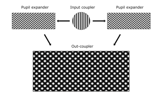
In this work we propose several full-color metagrating solutions for single waveguide-based
Augmented and Virtual Reality near-eye display systems. The presented solutions are based on a combination of
reflective and/or transmissive diffraction gratings inside or outside a waveguide. The proposed in-coupler
designs have high diffraction efficiency across a wide angular range. Applying our new grating combination
solution, we can provide good gathering of diffracted rays for the different colors. We demonstrate that by using
a dual-mode symmetrical in-coupling system and angular pupil tiling, we can extend the overall horizontal FoV
for three RGB colors. The new characteristics of the full single waveguide system including Eye Pupil Expander
and out-coupling components compatible with the proposed in-coupling solutions are discussed. We show that a
new nonsymmetrical design of metagratings can be used to change its diffraction properties improving the
diffraction efficiency and diffraction uniformity of the optical components.
“Metagrating solutions for full color single-plate waveguide combiner” Oksana Shramkova * , Valter Drazic, Guillaume Bourcin, Bobin Varghese, Laurent Blondé, and Valérie Allié , Metamaterials for Novel Wave Phenomena in Microwaves, Optics, and Mechanics. 2022.
Waveguide based optical combiners for augmented reality (AR) glasses are integrating several surface relief gratings (SRG) whose pitch sizes can be as small as 200 nm for the blue wavelength. All SRG components exploit the first diffraction order to couple in and out or to deviate the light. We present SRG using higher diffraction orders featuring over-wavelength pitch sizes. Our gratings use the edge wave (EW) diffraction phenomenon to steer light in the
preferred far field direction.
“Over-wavelength pitch sized diffraction gratings for augmented reality applications .“, Drazic, Valter; Shramkova, Oksana; Varghese, Bobin; Blondé, Laurent; De La Perrière, Vincent Brac; Schiffler, Jesse; Twardowski, Patrice; Lecler, Sylvain; Walter, Benjamin; Mairiaux, Estelle; Bavedila, Fuanki; Faucher, Marc; Allié, Valérie. 2022 Optics Express 30(2) 1293-1303
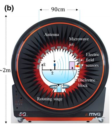
In this paper, we report the experimental and numerical investigation of plane wave diffraction by an all-dielectric dual-material cuboid. Edge diffraction by a cuboid leads to the generation of a narrow, high intensity beam in the near-field region called a photonic jet. We examine the dependence of the jet behavior and orientation on the materials and dimensions of constitutive parts in the microwave frequency domain. The possibility to shift and deviate the resultant microwave jet in the near-field region of such a structure depending on the size of constitutive parts is demonstrated numerically. Experimentally, we observe a shift in the spatial position of the jet. The experimental asymmetric electric field profile observed in the far-field region is attributed to the input of multiple edge waves generated by the dual-material cuboid. The presented results may be scaled at different frequency bands such as optical frequencies for designing nanostructures enabling the focusing and deviation functionality and creation of new optical devices which would satisfy the needs of emerging nanophotonic applications.
“Experimental observation of asymmetrical microwave jets and far-field distribution generated by a dual-material system“, B. Varghese1, , O. Shramkova1, P. Minard2, L. Blondé1, V. Drazic1, V. Allié1, Scientific reports, june 2021.
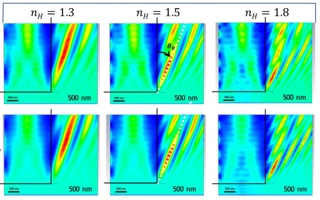
Spherical microparticles have the ability for nonresonant focusing of light in the near field zone, forming nanojet (NJ) beams. Arbitrary-shaped microstructures, with wave-length-scale dimensions, may offer similar functionality with lower fabrication complexity. The focusing properties are ruled by the edge diffraction phenomenon. The diffraction of light on the edge of a dielectric microstructure forms a tilted focused beam whose deviation angle depends on the index ratio between the structure material and host medium. The beam geometry and field intensity enhancement can be tuned by varying the curvature of the edge line. Interference of edge diffracted waves from different segments of the edge line creates a condensed beam in the nearfield zone, the photonic nanojet.
“Near field focusing by edge diffraction“, A. Boriskin, V. Drazic, R. Keating, M. Damghanian, O. Shramkova, L. Blondé. Optics Letters, vol. 43, Issue 16, pp. 4053-4056 (2018)
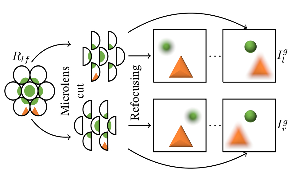
 In this paper, we present a complete processing pipeline for focused plenoptic cameras. In particular, we propose (i) a new algorithm for microlens center calibration fully in the
Fourier domain, (ii) a novel algorithm for depth map computation using a stereo focal stack and (iii) a depth-based rendering algorithm that is able to refocus at a particular depth or to create all-in-focus images. The proposed algorithms are fast, accurate and do not need to generate Subaperture Images (SAIs) or Epipolar Plane Images (EPIs) which is capital for focused plenoptic cameras. Also, the resolution of the resulting depth map is the same as the rendered image. We show results of our
pipeline on the Georgiev’s dataset and real images captured with different Raytrix cameras.
“An Image Rendering Pipeline for Focused Plenoptic Cameras“, M. Hog, N. Sabater, B. Vandame, V. Drazic, IEEE Transactions on Computational Imaging, Vol. 14, No 8, August 2015.
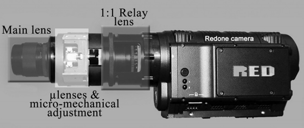
 A plenoptic camera is a natural multiview acquisition device also capable of measuring distances by correlating a set of images acquired under different parallaxes. Its single lens and single
sensor architecture have two downsides: limited resolution and limited depth sensitivity. As a first step and in order to circumvent those shortcomings, we investigated how the basic design parameters of a plenoptic camera optimize both the resolution of each view and its depth-measuring capability. In a second step, we built a prototype based on a very high resolution Red One® movie camera with an
external plenoptic adapter and a relay lens. The prototype delivered five video views of 820 × 410. The main limitation in our prototype is view crosstalk due to optical aberrations that reduce the depth accuracy performance. We simulated some limiting optical aberrations and predicted their impact on the performance of the camera. In addition, we developed adjustment protocols based on a simple pattern and analysis of programs that investigated the view mapping and amount of parallax crosstalk on the sensor on a pixel basis. The results of these developments enabled us to adjust the lenslet array with a submicrometer precision and to mark the pixels of the sensor where the views do not register properly.
“Optimal design and critical analysis of a high-resolution video plenoptic demonstrator“, V. Drazic, JJ Sacré, A. Schubert, J. Bertrand, E. Blondé. Journal of Electronic Imaging 21(1), 011007 (Jan–Mar 2012) .
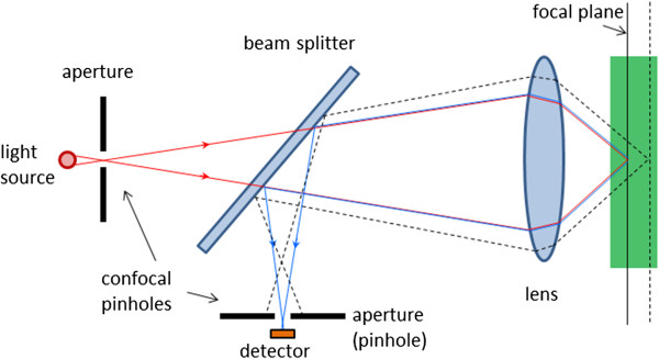
We consider the effect of the finite size of the source and of the detector on the three-dimensional transfer function of an incident light coherent confocal scanning microscope. Up to now the source has always been modeled as being punctiform. We will show that a finite source alters the 3D resolution and hence the imaging properties of such a microscope much more than a finite detector of the same size. It is also the aim of this paper to determine the greatest size of the source and the detector which still preserves the 3D resolution attained with a confocal microscope.
“Three-dimensional transfer function of coherent confocal microscopes with extended source and detector“, Valter DRAZIC, Journal of Modern Optics, 1992, vol. 39, No. 8, 1777-1790.
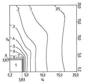
Previous papers about coherent scanning optical microscopes took into account two types of microscope: those with a point detector called type-II or confocal microscopes and those with an infinitely large area detector called type-I or conventional microscopes. Here the pinhole size of a type-II microscope was permitted to vary, and it is shown how the size could affect the imaging properties of a real microscope. The three-dimensional optical transfer function is established, and we discuss in particular the resolution capabilities, lateral as well as longitudinal, of a scanning microscope with a given pinhole size or detector area. Finally, a rigorous confocality criterion, which will answer the question of how small the pinhole should be made to give confocal imaging properties to a scanning microscope, is given.
“Dependence of two- and three-dimensional optical transfer functions on pinhole radius in a coherent confocal microscope“, Valter DRAZIC, Journal of the Optical Society of America A, Vol. 9, No 5, May 1992, 725-731
“Dependence of two- and three-dimensional optical transfer functions on pinhole radius in a coherent confocal microscope – comments“, C. J. R. Sheppard, Min Gu, Journal of the Optical Society of America A, Vol. 10, No 3, March 1993, 533-534
“Dependence of two- and three-dimensional optical transfer functions on pinhole radius in a coherent confocal microscope – reply to comments“, Valter DRAZIC, Journal of the Optical Society of America A, Vol. 10, No 3, March 1993, 535-537